Figure 3.
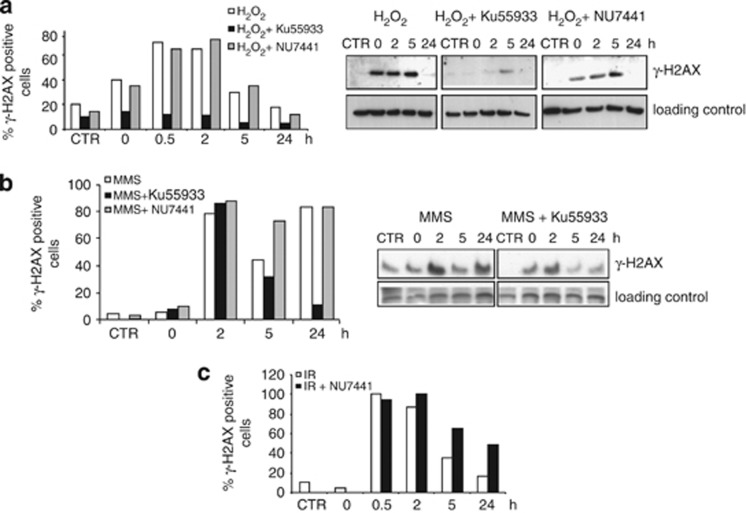
γH2AX response is activated in myotubes upon SSB induction and ATM is the main kinase involved. Induction of γH2AX foci after H2O2 (a), MMS (b) or IR (c) treatment of terminally differentiated muscle cells. Myotubes were exposed to the DNA-damaging agent with or without 1 h pre-treatment with a specific kinase inhibitor (KU55933 or NU7441), and analyzed at different times after damage. (a) DNA-damage induction and repair as detected by γH2AX foci formation (left) or western blotting (right) after exposure to H2O2 (100 μM, 30 min). (b) DNA-damage induction and repair as detected by γH2AX foci formation (left) or western blotting (right) after exposure to MMS (3 mM, 30 min). (c) DNA damage induction and repair as detected by γH2AX foci formation after exposure to IR (2 Gy). Experiments were repeated twice and one representative experiment is shown. In the γH2AX foci assay, at least 200 nuclei were examined for each time point. Western blot analysis was performed on nuclear cell extracts, and the loading control is DNA polymerase β. CTR, control cells untreated

