
Figure 1.
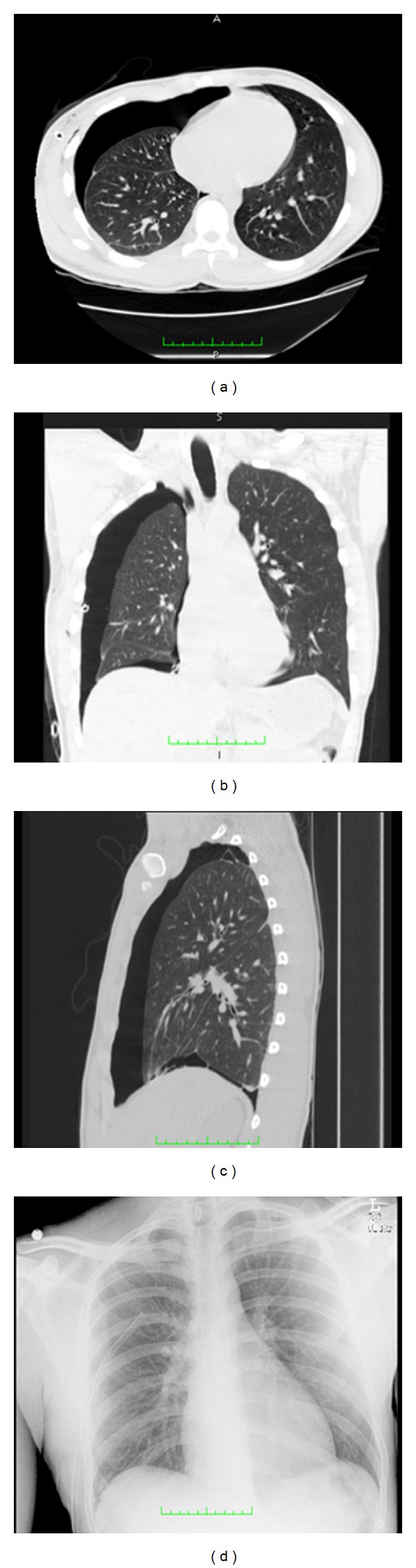
Transverse (a), coronal (b), and sagital (c) CT images of a patient with a moderate to large pneumothorax, and chest X-ray (d) of the same patient taken two hours prior to the time the CT images were acquired. CT images provide significantly better detail for detection and quantification of pneumothorax size.