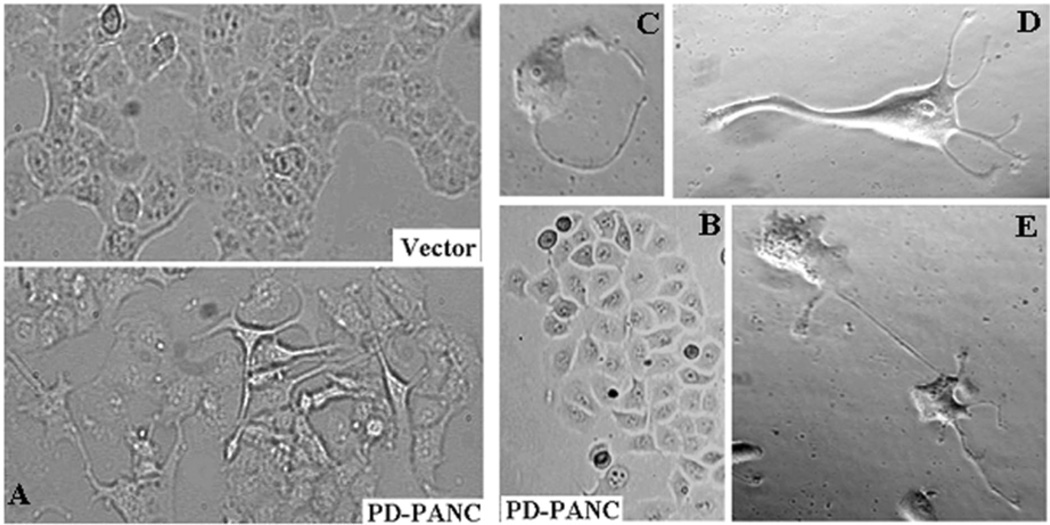
Figure 1.
Morphology of PD-PANC-1s. A-Vector-controls (Left-top) or PD-PANC-1s (Left-bottom) were plated at high density. Note cells with long filopodia present in the PD-PANC-1s culture that are not present in Vector-controls (X4 objective). B- PD-PANC-1 culture displaying epithelial cuboidal morphology (X4 objective). C-E-micrographs (X20 phase objective) of low density plated PD-PANC-1s showing large individual cells with filopodia.

