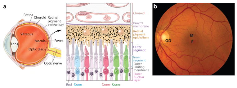
Figure 1.
(a) Cross-section of the human eye, and schematic of the photoreceptor-retinal pigment epithelium (RPE)-choroidal layers that are affected in AMD. Cone photoreceptors are shown in red, green, or blue. RPE apical processes are intimately associated with photoreceptor outer segments. Two of the rods are shown in the active process of outer segment disc shedding. Melanosomes are shown as dark organelles in RPE cells. (b) Fundus photograph showing the retina of a normal individual. Retinal blood vessels are clearly visible. F, fovea; M, macula; OD, optic disc.

