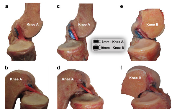
Figure 3.
Physiologic and reconstructed ACL movement pattern. A medial view of the native ACL of knee A in full extension (a) and flexion (b). The two bundles are roughly parallel in extension and cross in flexion. A medial view of the same knee following ACL reconstruction utilizing oval drill holes and rectangular bone blocks in full extension (c) and flexion (d). The graft behavior is qualitatively the same as the native ligament. A medial view of knee B following reconstruction using standard bone blocks and tunnels is shown in full extension (e) and flexion (f). Again the graft behavior is qualitatively the same as the native ligament. The schematic drawing shows the relation between bone blocks and tunnels in knee A and B.

