
Figure 2.
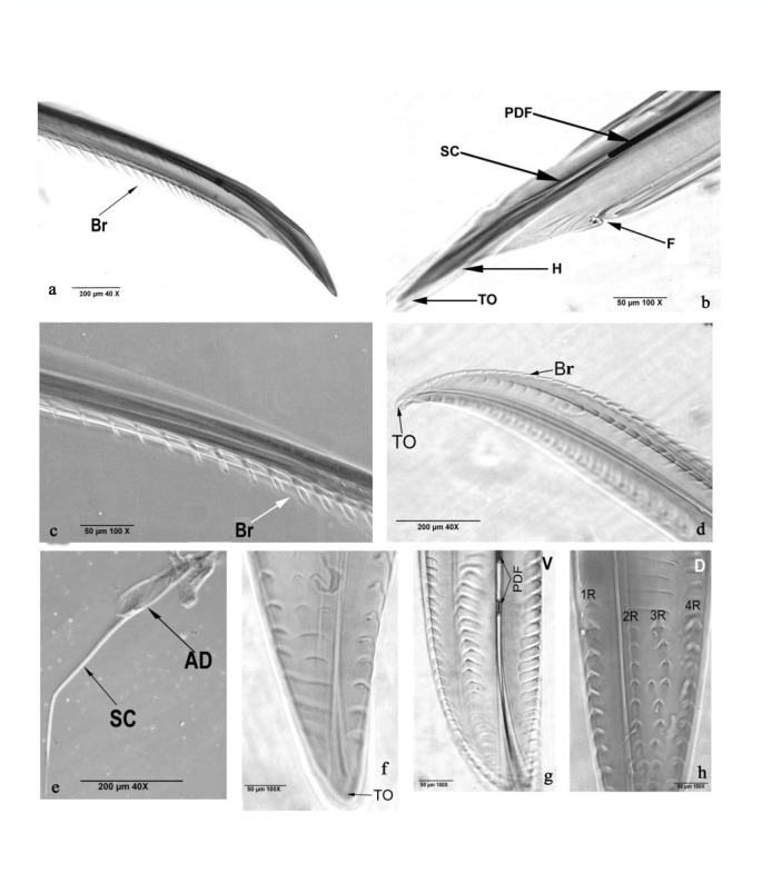
Rhynocoris marginatus phase contrast photographs of lateral (a and b) and terminal (c) view of maxillary stylet, magnified tip region of mandibular stylet (d), terminal region with adductor muscle (e), ventral (f and g) dorsal (h) view of mandibular tip. AM adductor muscles, Br - brush-like, F - furrow, H - hook PDF partially digested food material, 1R - first row, 2R - second row, 3R - third row and 4R - fourth row. SC - salivary canal, TO - terminal opening. High quality figures are available online.