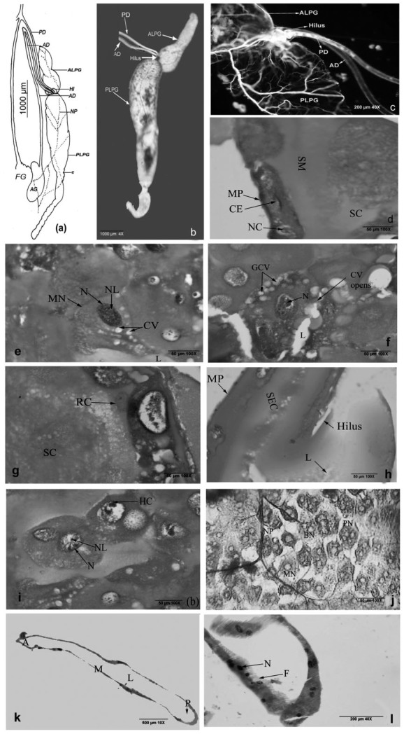
Figure 3.

Gross morphology of Rhynocoris marginatus salivary gland - line diagram (a) principle gland (4×) (b), hilus region (c), anterior part of the posterior lobe (d), secreting cells of posterior lobe (e), collecting vacuoles of the posterior lobe (f), replacement cells and secretions (g), hilus cross section (h), posterior lobe double nucleated cells (i) principal gland surface cells (j), longitudinal section of accessory gland (k), lower portion of accessory gland (I). (A anterior region, ALPG -anterior lobe of principle gland, AD accessory duct, AG - accessory gland, BN - binucleated, CE - cubic epithelium, CV opens- collecting vacuoles opens, GCV - group of collecting vacuoles, HI- hilus, F-filament, HC- heterochromatin, LLumen, M- mid region, MN- mononucleated, MP- membrane probia, N - nucleus, NL - nucleolus, NP - nerve plexus, P- posterior region, PLPG - posterior lobe of principle gland, PN - polynucleated, SC secretory epithelium, Sc - secretions in dense, SM - secretory materials TN - tirnucleated, T - trachea, To - tracheoles). High quality figures are available online.
