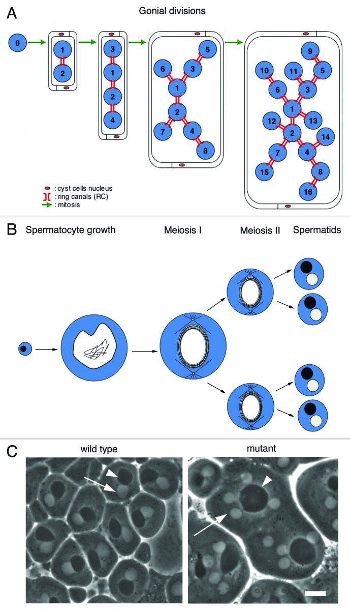
Figure 1. Spermatogenesis in Drosophila melanogaster. (A, B) Schematic representation of spermatogenesis in D. melanogaster. (A) A single primary spermatogonium undergoes four mitotic divisions. Numbers indicate gonial cells, number 0 indicates the primary spermatogonium, the mitotic founder of a cluster of dividing secondary spermatogonia connected by ring canals. Two cyst cells engulf the progeny of the primary spermatogonium throughout spermatogenesis. Blue, spermatogonia. (B) Each primary spermatocyte undergoes a growth phase, which lasts 90 h before undergoing two meiotic divisions. A wild type spermatid cyst at the so called onion stage contains 64 spermatids connected by 63 ring canals (not shown). Each spermatid contains a single nucleus (white) associated with a nebenkern (black) of similar size. Only four spermatids are represented. Blue, cytoplasm. (C) Spermatids at the onion-stage viewed by phase-contrast microscopy. Each wild type spermatid contains a single light nucleus (arrow) associated with a dark nebenkern (arrowhead). Spermatids from mutants defective in male meiotic cytokinesis, contain large nebenkerne (arrowhead) associated with 2 or 4 nuclei of similar size (arrow). Bar, 10μm.

An official website of the United States government
Here's how you know
Official websites use .gov
A
.gov website belongs to an official
government organization in the United States.
Secure .gov websites use HTTPS
A lock (
) or https:// means you've safely
connected to the .gov website. Share sensitive
information only on official, secure websites.
