FIGURE 1:

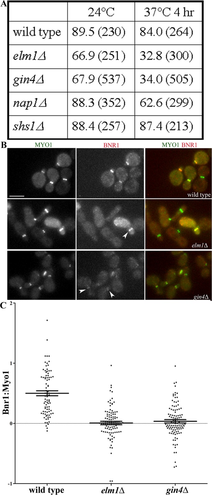
Partial requirement for Gin4 and Elm1 for the localization of full-length Bnr1. (A) The absence of Gin4 or Elm1 disrupts the localization of Bnr1-GFP to the bud neck, unlike the absence of Nap1 and Shs1. Shown is the mean percentage of Myo1-CFP–positive cells with Bnr1-GFP localization at the bud neck for the number of cells counted (indicated in parentheses). Experiments were performed twice with >100 cells per sample. (B) Representative images of Bnr1-GFP and Myo1-CFP in the indicated strains. Cells were fixed after 4 h at 37°C and imaged in three dimensions with five 0.5-μm stacks using the same exposure conditions for all strains. Scale bar, 5 μm. Shown are representative, normalized, maximum-projection images. (C) The ratio of the fluorescence intensity of Bnr1 (YFP) to Myo1 (CFP) was measured (see Materials and Methods for details) in the cells imaged in B. Each point represents the ratio of the background-corrected fluorescence intensity of Bnr1 to Myo1 for one cell; bars, mean ± SE.
