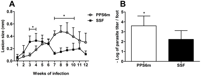
Figure 6. Isolate with high ecto-nucleotidase activity shows delay in lesion development in C57BL/6J mice.
C57BL/6J mice were inoculated in the footpad of the left hind leg with 107 promastigotes (A). Lesion sizes were measured weekly. Data-points represent the mean+SD of the difference between the infected and uninfected contralateral footpad from two independent experiments with four mice per group. (B) Parasite load in lesions. Twelve weeks after infection, animals were sacrificed and the parasite load determined by limiting dilution technique in the infected footpad. Columns represent the mean+SD of the log parasite titer from the lesions of four mice per group from two independents experiments. (*) indicates statistical difference between isolates. Statistical analysis was performed by two-way ANOVA followed by Bonferroni post-test (A) and Students's t-test (B).

