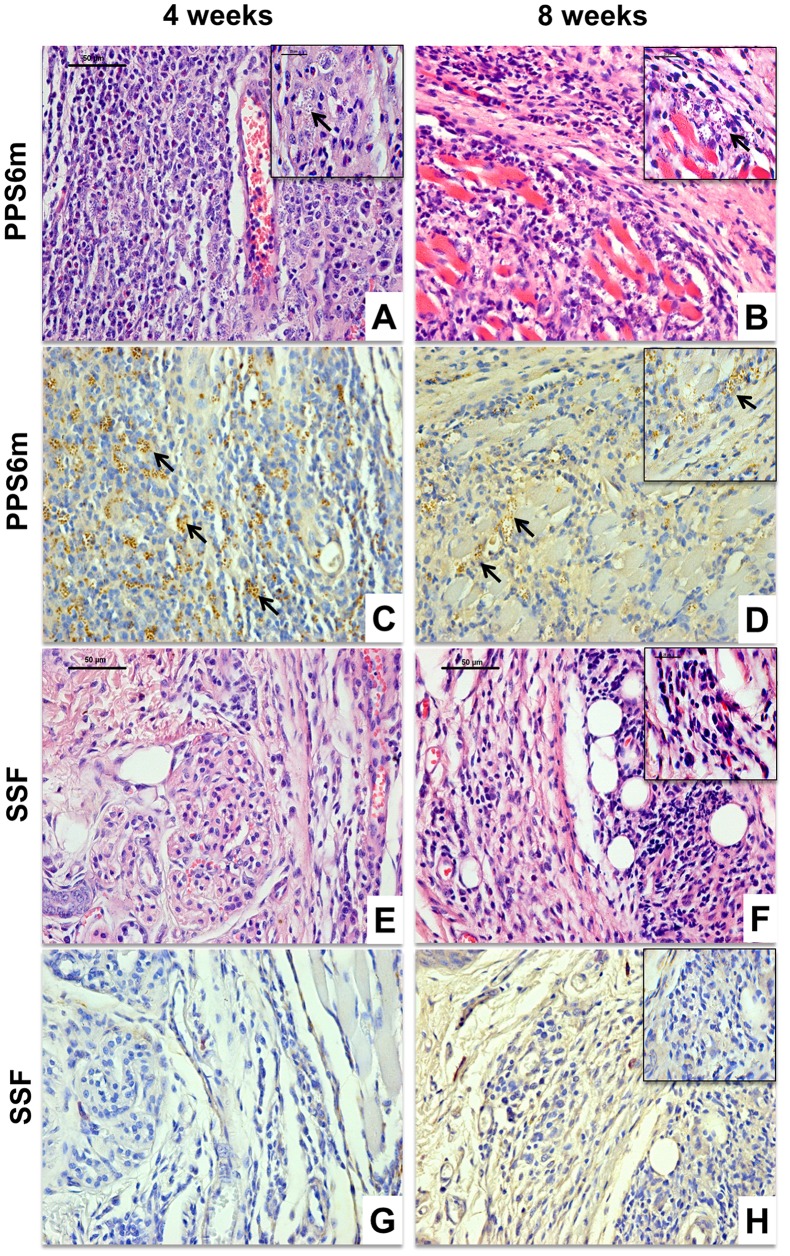
Figure 7. Histological (4 µm, HE) and immunohistochemical evaluation of C57BL/6J mice infected by L. (V.) braziliensis isolates.
C57BL/6J mice were inoculated in the footpad of the left hind leg with 107 promastigotes from PPS6m (A–D) and SSF (E–H) isolates for 4 (A, C, E and G) and 8 (B, D, F and H) weeks. Bar = 50 µm, 40×. Insert: Bar = 25 µm, 100×. Arrows indicate the presence of amastigotes. Figures are representative of the Histological (A, B, E and F) and immunohistochemical evaluation (C, D, G and H) of at least two independent experiments with four animals per group.

