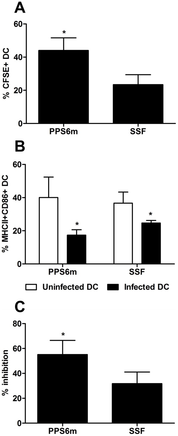
Figure 8. DC infection and inhibition of activation is increased in PPS6m isolate.

BMDC obtained after 9 days of culture with GM-CSF were infected with CFSE-labeled metacyclic promastigotes (3 parasites/cell). After 3 hr, the cells were stimulated with 2 µg/mL LPS and then incubated for up to 17 hr. Finally, DC were analyzed by flow cytometry. DC were gated into populations of uninfected (CFSE− cells) and infected (CFSE+ cells) BMDC, and the surface markers MHCII and CD86 analyzed in both populations. (A) Percentage of CFSE+ DC. (B) Percentage of MHCII+CD86+ DC. (C) Reduction of MHCII+CD86+ cells in the population of infected DC compared to uninfected DC. Bars represent the mean+SD of three independent experiments. (*) indicates statistical difference (p<0.05). Statistical analysis was performed by Students's t-test.
