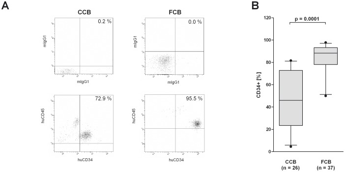
Figure 2. Differences and parallelism in the purity of CD34+ -cell-purifications from FCB and blood bank CCB.
(A) MNC were isolated from FCB and blood bank CCB by washing with DNAse buffer or Ficoll-paque density gradient centrifugation as described in materials and methods. CD34+ stem cells were isolated from MNC by a positive magnetic separation of CD34+ cells and the purity was analyzed by flow cytometry. Dot plots depict the percentage of CD34+ cells of one representative separation from CCB and FCB (lower panels). The quadrant was set according to the isotype controls (upper panels). (B) The bar chart depicts the percentage of CD34+ cells after isolation of CD34+ cells from 26 CCB and 37 FCB samples. Box plots depict median and 5–95% percentile. Level of significance is given. n. s. = no significance.

