Fig. 2.
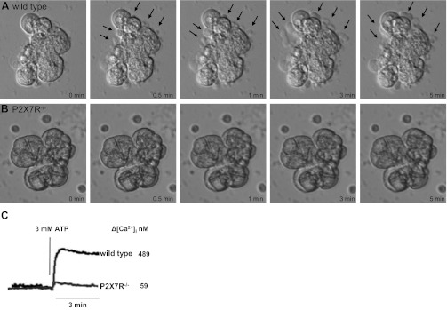
ATP induces membrane blebbing and increases in intracellular calcium concentration ([Ca2+]i) in SMG cells from wild-type, but not P2X7R−/−, mice. Freshly dispersed SMG cell aggregates from wild-type (A) or P2X7R−/− (B) mice were treated at pH 7 with 3 mM ATP, and cells were monitored by real-time brightfield microscopy for 5 min at 37°C. Select still-frames of cell micrographs are shown at the indicated time points. Membrane blebs (arrows) can be clearly seen within 30 s of ATP treatment in wild-type SMG cells and increase over 5 min. C: ATP-induced changes in [Ca2+]i in single SMG cells were quantified using the Ca2+-sensitive fluorescent dye fura-2 and an InCyt dual-wavelength fluorescence imaging system, as described in materials and methods, and the peak increase in [Ca2+]i for n = 9 cells is indicated. Images and [Ca2+]i traces are representative of results from three independent experiments.

