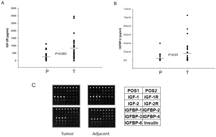
Figure 3. The differential expression of IGF-2R and IGFBP-2.
IGF-2R (A) and IGFBP-2 (B) were differentially expressed between tumor (T) and matching paratumorous (P) samples. Liver cancer tissue and adjacent tissue lysates from 25 patients was prepared and incubated with IGF signaling antibody arrays, and the data was statistically analyzed. The average concentrations of IGF-2R and IGFBP-2 expressed differentially from liver cancer and adjacent tissues were compared and the P<0.05 (T test). The representative data of Antibody Arrays are shown in (C).

