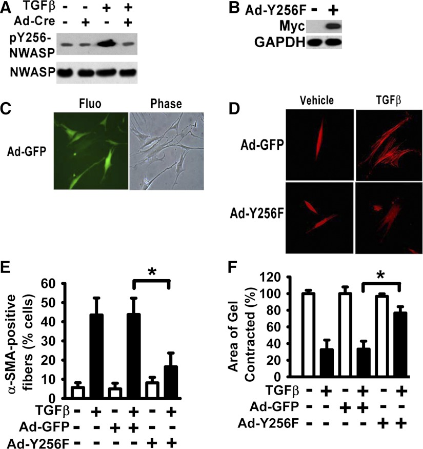
Fig. 4.
Expression of N-WASP Y256F mutant significantly inhibited the formation of α-SMA-containing filaments and cell contraction in lung fibroblasts treated with TGF-β1. A: lung fibroblasts derived from FAK-floxed mice were treated with or without Ad-Cre for 24 h and treated with or without TGF-β1 (10 ng/ml) in SFM with 1% BSA for 6 h (37°C, 5% CO2) as shown in Fig. 3. Cells were lysed, and equivalent amount of whole cell lysates were subjected to Western Blot analysis with indicated antibodies. B: human lung fibroblasts were infected with adenoviral vector containing the myc-tagged Y256F N-WASP mutant (Ad-Y256F), and equivalent amount of whole cell lysates were subjected to Western Blot analysis with indicated antibodies. C: human lung fibroblasts were infected with adenoviral vector containing the GFP (Ad-GFP) for 48 h. Fluorescent image for GFP expression (left), phase contrast image (right) (×200). D: lung fibroblasts were infected with Ad-Y256F or Ad-GFP, treated with or without TGF-β1 (10 ng/ml) for 48 h, fixed, and stained with Cy-3-labeled antibody toward α-SMA as shown in Fig. 1 (×200). E: percentage of cells with α-SMA-containing filaments in D was quantified as in Fig. 1. Data are presented as the means ± SE. *P < 0.01. F: lung fibroblasts were treated as in E and subjected to collagen gel contraction assay as shown in Fig. 2. *P < 0.01.

