Fig. 4.

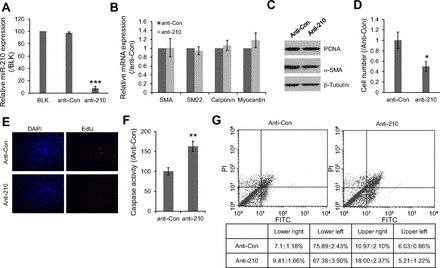
Functions of hypoxia-induced miR-210 in HPASMC. A: expression level of miR-210 in hypoxic HPASMC after transfection with either miR-210 knockdown probe or control probe (anti-Con) were investigated by qPCR. The values are shown relative to the value obtained from blank control. B: mRNA expression levels of α-smooth muscle actin (SMA), SM22, Calponin, and myocardin were examined by qPCR from the same samples mentioned above. 18S rRNA was used as reference. Data was shown relative to control probe. C: protein levels of PCNA and SMA were also examined in the treated cells. β-Tubulin was used as an internal control. D: effect of miR-210 knockdown on cell growth was evaluated by counting the cell number. The values are shown relative to the value obtained with control probe. E: effect of miR-210 knockdown on cell proliferation was also evaluated by EdU incorporation assay. F: induction of apoptosis due to the loss of miR-210 in hypoxic HPASMC was evaluated by measuring Caspase-3/7 activities. G: effects of miR-210 knockdown on cell apoptosis at hypoxia condition were also investigated by analyzing cell death with FACScan flow cytometer. Cells were stained with FITC-conjugated annexin V and PI. All the values are shown relative to the value obtained with control probe. *P < 0.05, **P < 0.01, ***P < 0.001.
