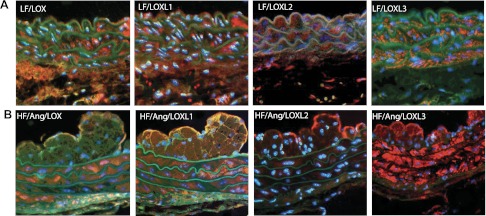
Fig. 3.
Localization of LOX/LOXL proteins in aortic tissue from ApoE−/− mice after 8 wk. A: localization of LOX/LOXL family members in aortic tissues of ApoE−/− mice after treatment with a low-fat diet for 8 wk. The spatial distribution is similar to that of the high-fat (HF) + ANG II treatment animals, with the exception of the neointimal space, which is not detected in low-fat fed animals after 8 wk. B: localization of LOX/LOXL family members in aortic tissues of ApoE−/− mice after treatment with a high-fat diet + ANG II for 8 wk. Protein expression was concentrated in the media, neointimal, and endothelial layers for LOXL1 and LOXL3. Expression of LOX was present in the medial layer and appears in the endothelial cell layer, but is absent from the neotintima. LOXL2 staining was present most abundantly within the endothelial cell layer.

