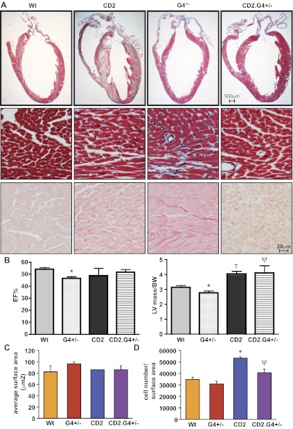
Fig. 4.
CD2 rescues G4+/− haplo-insufficient hearts. A: trichrome staining (top and middle) and Sirius red staining (bottom) of whole heart sections from adult WT, HA-CD2 transgenic, G4+/−, and CD2.G4+/− mice (170 days old). Note the hypoplastic heart of G4+/− mice and the larger LV of the CD2 mice. Note as well the collagen deposition (blue in trichrome and red in Sirius red staining) in the G4+/− heart as evidence of fibrosis. B: echocardiography analysis showing the LV mass measurement corrected by the body weight (LV/BW) and the EF% of the different mouse genotypes; N = 5–6 for each group. C: average cell surface area calculated from ventricular sections of adult WT, CD2 transgenic, G4+/−, and CD2.G4+/− mice (170 days old). The histogram represents quantification of 5 fields from 2 different hearts for each group. The results presented are means ± SE. D: total cell number in ventricular and interventricular septum sections of adult WT, CD2 transgenic, G4+/−, and CD2.G4+/− mice (170 days old). The results presented are means ± SE; n = 3 for each group. *P < 0.05 vs. WT; τP < 0.005 vs. WT; ΨP < 0.05 vs. G4+/−.

