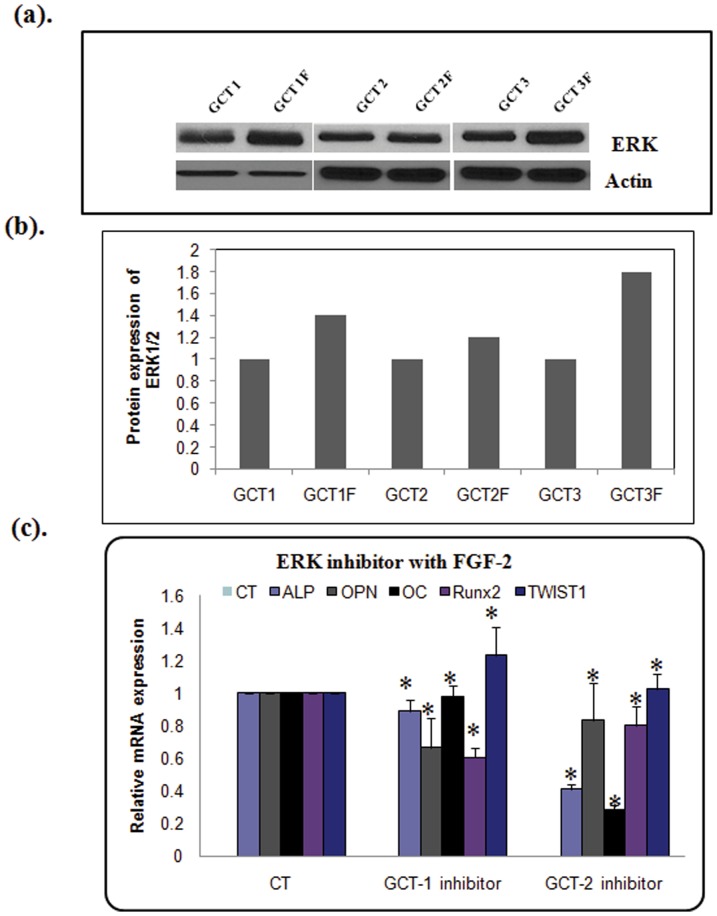
Figure 7. FGF2 signaling through ERK in osteoblast differentiation in GCT stromal cells.
(a) Western blot analysis (b) and quantification showed that FGF-2 stimulation resulted in increased phosphorylation of ERK1/2 (p-ERK1/2), compared to untreated GCT cells. β-Actin was used as loading control. (c). Inhibition of osteoblastic gene expression by ERK inhibitor in GCT stromal cells based on real-time RT-PCR. GCT stromal cells were treated with 1 nM ERK-inhibitor PD98059 (all diluted in DMSO) for 24 h in serum-free medium. mRNAs were purified, cDNAs were synthesized, and the samples were analyzed by real-time PCR. The ΔΔCT method was used to calculate the real-time RT-PCR fold change using RPS18 mRNA as an endogenous control, and all changes in expression are relative to the control without treatment. Three independent real-time PCR runs were performed on each sample.

