Fig. 1.
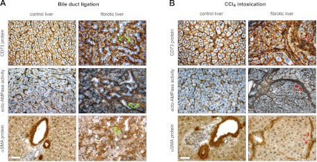
CD73 protein expression and enzymatic activity redistribute to myofibroblast-rich regions in experimental liver fibrosis. A: effects of bile duct ligation (BDL) on CD73 expression and activity. Liver sections from rats subjected to BDL (2 wk) were used to visualize in situ ecto-AMPase activity, by the Wachstein/Meisel lead phosphate precipitation method, and ecto-5′-nucleotidase/CD73 and α-smooth muscle actin (α-SMA) proteins, by using a standard peroxidase-based immunohistochemistry procedure. In control animals, both ecto-AMPase activity and ecto-5′-nucleotidase/CD73 protein expression have parallel localization at the level of both canalicular and sinusoidal membrane domains of hepatocytes and the hepatic portal areas, whereas α-SMA protein is only observed at the level of the smooth muscle cell layer surrounding blood vessels. Upon hepatic fibrosis induction, ecto-AMPase activity and ecto-5′-nucleotidase/CD73 and α-SMA protein expression are seen in the vicinity of fibrotic areas surrounding proliferating bile ducts (green arrows). B: effects of CCl4 intoxication on CD73 expression and activity. The effects of CCl4-induced liver fibrosis are even more profound, as the distribution of ecto-5′-nucleotidase/CD73 protein expression and activity shifted to broad fibrotic bands surrounding hepatocyte regenerative nodules in the same locale as α-SMA-positive liver myofibroblasts (red arrows). Scale bar, 40 μM.

