Fig. 5.
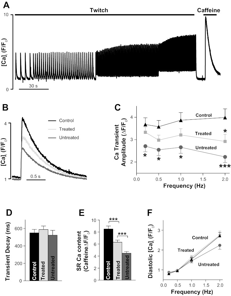
APAU treatment partially prevents the decrease in Ca2+ transient amplitude and sarcoplasmic reticulum (SR) Ca2+ content in UCD-T2DM rats. A: representative example of Ca2+ transient and SR Ca2+ content measurements. Fluo-4 -loaded cardiac myocytes were paced with external electrodes at 0.2, 0.5, 1, and 2 Hz until Ca2+ transients reached steady state. After 2 Hz, pacing was stopped for 10 s, and we applied 10 mM caffeine to measure SR Ca2+ content. B: representative examples of Ca2+ transients in myocytes from nondiabetic control rats, untreated UCD-T2DM rats, and UCD-T2DM rats treated with APAU. Myocytes were paced at 0.5 Hz. C–F: mean Ca2+ transient amplitude (C), Ca2+ transient decay time (D), SR Ca2+ content (E), and diastolic [Ca2+]i in cardiac myocytes from nondiabetic control rats, untreated UCD-T2DM rats, and UCD-T2DM rats treated with APAU. n > 20 myocytes from 4 different animals/group. Measurements were done at the end of the 6-wk treatment period. *P < 0.05 and ***P < 0.001 vs. the control group unless otherwise indicated.

