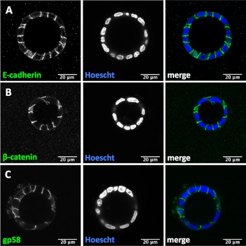
Fig. 3.
Localization of basolateral membrane markers in three-dimensional WT MDCK cell cysts. Immunofluorescence analysis of representative WT MDCK cell cysts demonstrates the localization of E-cadherin (A), β-catenin (B), and gp58 (C; green) at the basolateral membrane. Hoescht (blue) was used to label nuclei. Merged color images are depicted at right. All scale bars depicted are 20 μm.

