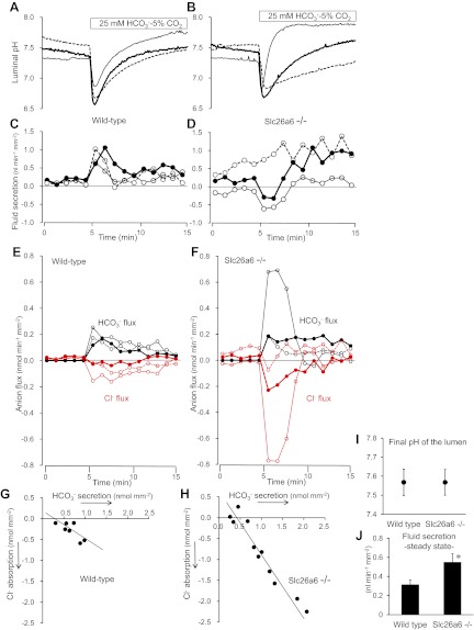
Fig. 1.
Cl−-dependent HCO3− and fluid secretion in interlobular pancreatic ducts isolated from wild-type and Slc26a6−/− mice. Sealed, isolated ducts from wild-type and Slc26a6−/− mice were filled with Cl−-rich (149 mM) HCO3−-free HEPES-buffered solution containing 2′7′-bis(2-carboxyethyl)-5(6)-carboxyfluorescein (BCECF)-dextran (20 μM). The bath was first perfused with the same HCO3−-free solution, and then the bath solution was switched to the standard HCO3−-buffered solution containing 25 mM HCO3− and 124 mM Cl− as indicated. Cells were stimulated with forskolin (1 μM) throughout the experiments. Luminal pH and luminal volume were measured simultaneously. A and B: changes in luminal pH in 3 representative ducts isolated from wild-type (A) and Slc26a6−/− (B) mice respectively. C and D: changes in fluid secretory rate in the same wild-type (C) and Slc26a6−/− (D) ducts. Negative values indicate fluid absorption. E and F: changes in calculated fluxes of Cl− (red) and HCO3− (black) in the same wild-type (E) and Slc26a6−/− (F) ducts. Positive and negative values indicate secretory and absorptive fluxes, respectively. G and H: plot of cumulative fluxes of Cl− and HCO3− during the first 5 min after bath application of HCO3− in wild-type (G, n = 7) and Slc26a6−/− ducts (H, n = 11). I and J: final values of luminal pH (I) and steady-state fluid secretory rate (J) in wild-type and Slc26a6−/− ducts. Data are means ± SE. *P < 0.05, significant difference.

