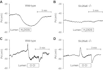
Fig. 6.
Effects of luminal H2DIDS and Cl− removal on membrane potential in wild-type and Slc26a6−/− ducts luminally-perfused with high Cl− solution. Bath and lumen were perfused with the standard HCO3−-buffered solution containing 25 mM HCO3− and 124 mM Cl−. Cells were stimulated with forskolin. Membrane potential (Pd) was measured by conventional microelectrodes. A and B: H2DIDS (200 μM) was applied to the lumen as indicated in wild-type (A) and Slc26a6−/− (B) ducts. Representative of 4 experiments. C and D: Cl− in the luminal solution was replaced with glucuronate as indicated in wild-type (C) and Slc26a6−/− (D) ducts. Representative of 4 experiments.

