Fig. 4.
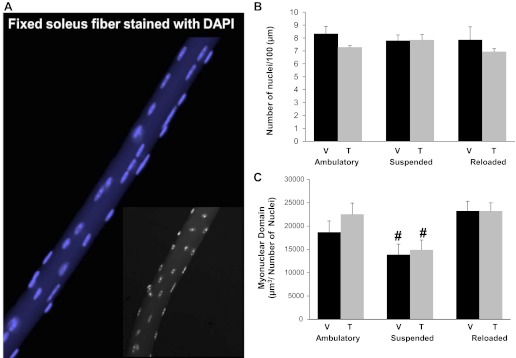
Myonuclear number remained constant across all experimental conditions, whereas myonuclear domain was reduced following suspension regardless of satellite cell depletion. A: representative image of an isolated fiber from a soleus muscle stained with DAPI (blue) to identify myonuclei. Inset: black and white image of an isolated fiber. B: myonuclear number was assessed in isolated soleus fibers. Nuclei were counted and represented as myonuclei per 100 μm of fiber length. C: myonuclear domain was determined by calculating the volume of a fiber segment (μm3) and normalizing to the number of myonuclei within the given segment. Values are presented as means ± SE. Black bars represent vehicle-treated animals and gray bars represent tamoxifen-treated animals. #Significant difference between treatment-matched ambulatory and suspended animals; P ≤ 0.05.

