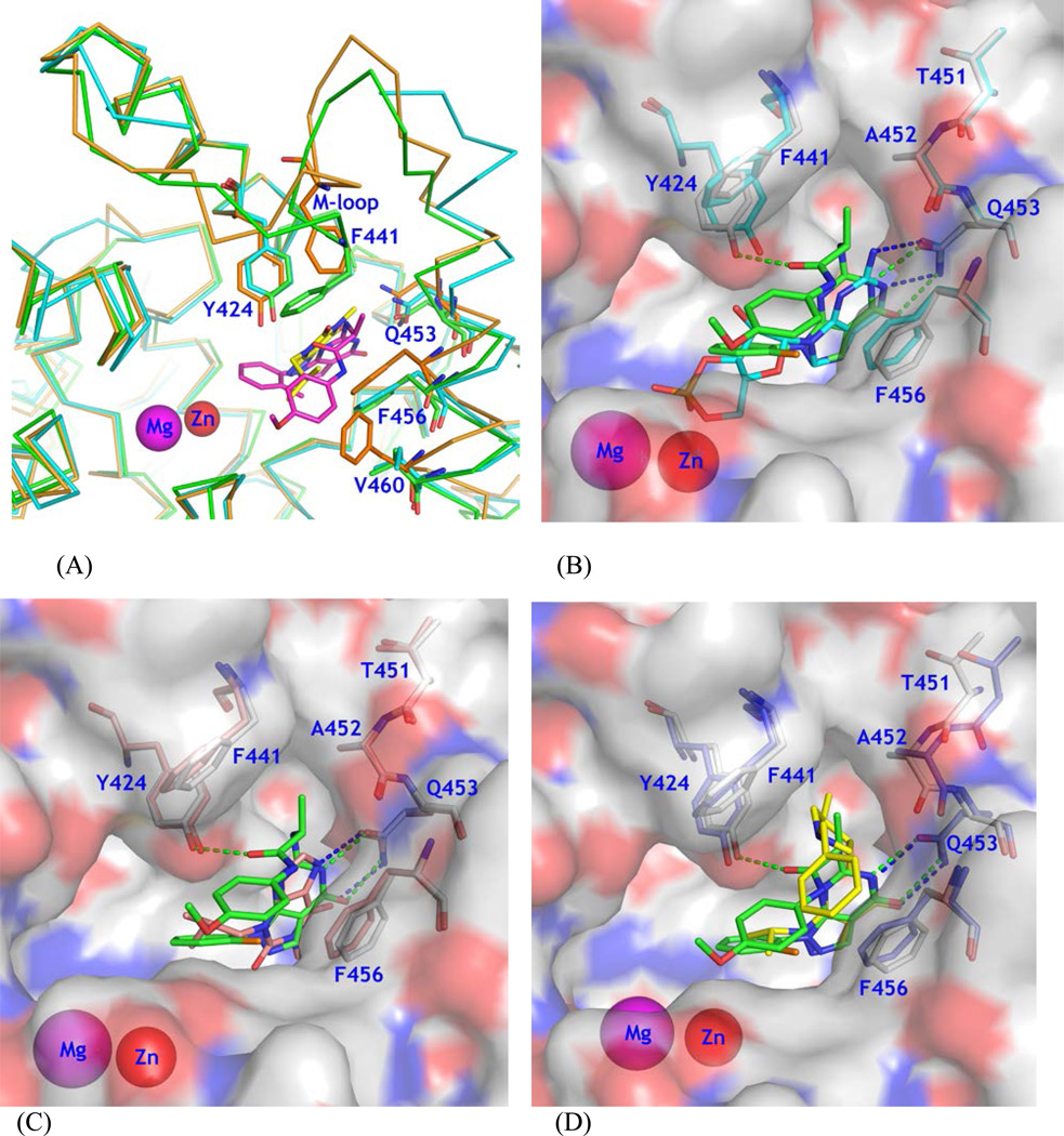
Figure 3.
Structural comparison. (A) Superposition of PDE9A (green sticks) over PDE1B (cyan), and PDE8A (orange). Inhibitor 28 of PDE9A is shown as purple sticks. IBMX of PDE8A is shown as yellow sticks. (B) A surface presentation for comparison on binding of 28 (green sticks) and cGMP (cyan) to the PDE9A active site. Residues were obtained from the superposition of the PDE9 complex structures. (C) Comparison on binding of 28 and IBMX (salmon). (D) Comparison on 28 and Pfizer inhibitor 7 (PF7, yellow). Residues Thr451 and Ala452 showed significant movement in the PDE9-PF7 structure.

