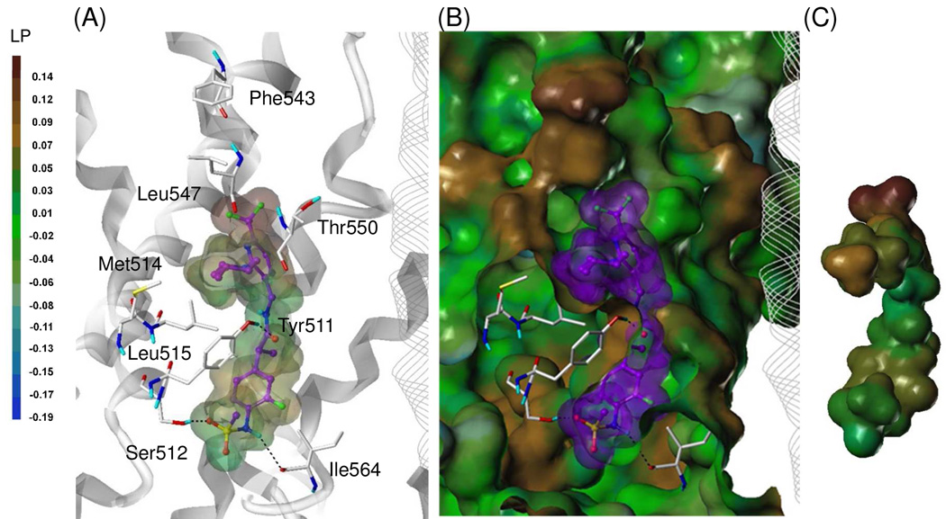
Figure 6.
Flexible docking of 49S in the hTRPV1 model.
(A) Binding mode of 49S. (B) Surface representations of the docked ligand and hTRPV1. (C) Van dar Waals surface of the ligand colored by its lipophilic potential property. The ligand is depicted as ball-and-stick with carbon atoms in purple; the details are the same as in Figure 5.

