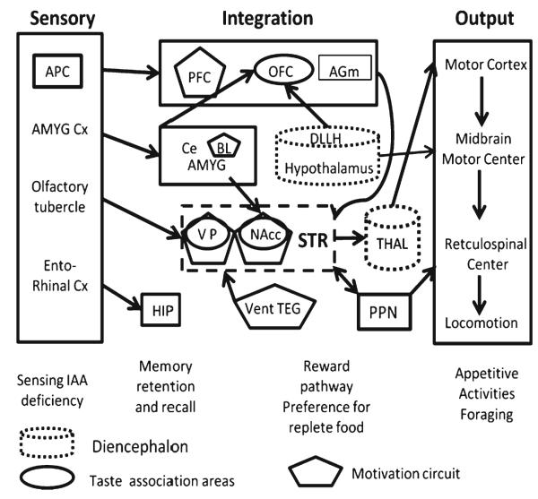
Fig. 3.
The brain areas associated with the sensory, integrative, and motor output functions associated with the sensing of and responses to IAA-deficient diets. Under Sensory, the regions belonging to the olfactory cortex, with their projections, are shown on the left of the figure. Under Integration, the top box represents cortical areas, prefrontal (PFC), orbitofrontal (OFC), and supplementary motor (AGm). As subcortical structures, central and basolateral amygdala (Ce, BL AMYG) and DLLH hypothalamus are as in Fig. 1. In the dashed box, ventral pallidum (VP) and nucleus accumbens (NAcc) are members of the basal ganglia in the striatum (STR). The ventral tegmentum (vent TEG) projects to the STR in the motivation circuit (Reward); at this level the pedunculopontine nucleus (PPN) also receives input from the STR and projects to reticulospinal centers. Members of the motivation circuit are in five-sided boxes, taste association areas are in ovals. The diencephalon includes the hypothalamus (and DLLH) and thalamus (THAL), indicated in dotted cylinders, for comparison with the lamprey circuit. Under Output are the motor centers for appetitive activities such as foraging. The hippocampus (HIP) is important for spatial memory

