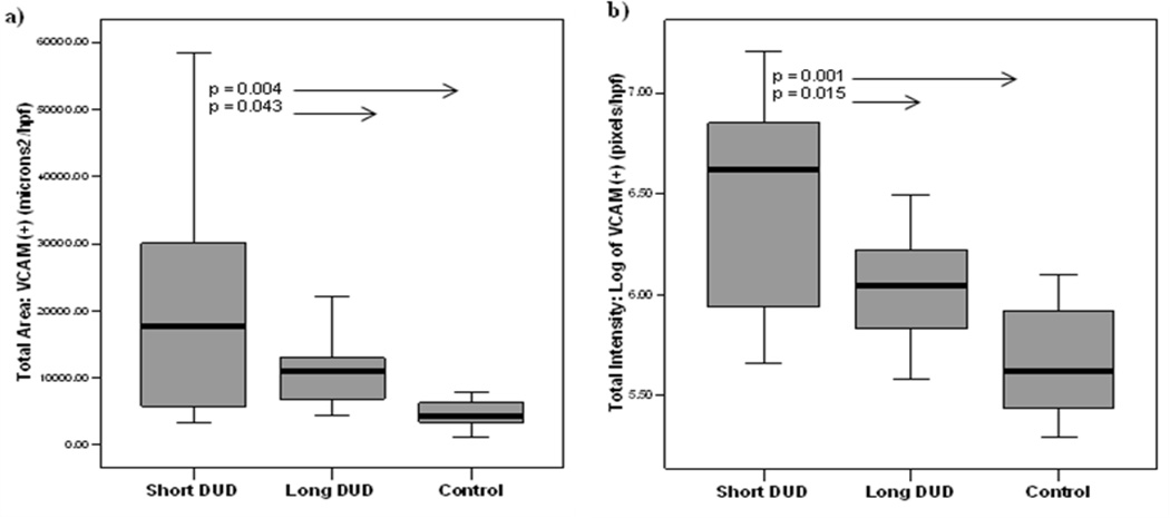
Figure 2. Increased total area and intensity of VCAM-1 positive staining in diagnostic muscle biopsies from untreated children with Juvenile Dermatomyositis of short disease duration.
a) Total area (microns²/hpf) and b) total intensity (pixels/hpf) of VCAM-1 positive expression in muscle biopsies increased in JDM short DUD compared with JDM of long DUD and controls. The rectangular box plot represents 50 percent of the data with the median value indicated by the line. The whiskers represent the smallest and largest values. JDM population: Short DUD, n=11 (less than 2 months), Long DUD (greater than 2 months), n=17 and age-matched controls, n=8.

