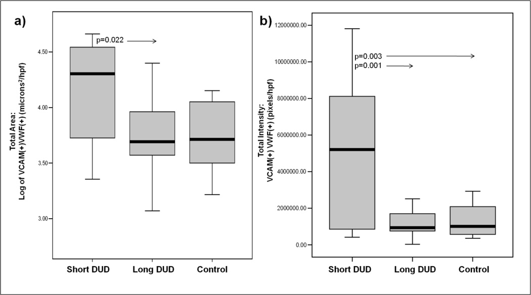
Figure 3. Increased VCAM-1 positive areas and intensity is localized to von Willebrand factor antigen positive vasculature in untreated JDM muscle biopsies of short disease duration.
a) Total area (microns²/hpf) and b) total intensity (pixels/hpf) of VCAM-1 positive and vWF positive expression in blood vessels of muscle biopsies of JDM short DUD (n=11), JDM long DUD (n=17) and controls (n=8). The rectangular box plot represents 50 percent of the data with median value indicated by the line. The whiskers represent the smallest and largest values. The additional circle outside the range indicates an outlier.

