Figure 3a.

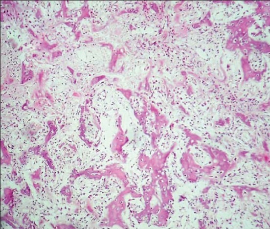
Photomicrograph of histopathologic section reveals irregular wove bone like tissue with osteoblastic rimming within fibrovascular connective tissue stroma. (H-E stain, × 100)

Photomicrograph of histopathologic section reveals irregular wove bone like tissue with osteoblastic rimming within fibrovascular connective tissue stroma. (H-E stain, × 100)