Figure 2.

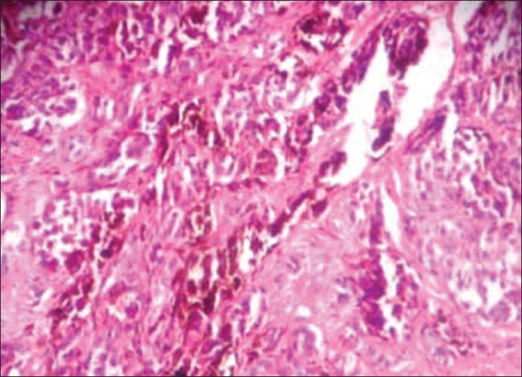
The hematoxylin and eosin–stained section shows melanoma with invasive pattern showing large cells with pleomorphic vesicular nucleus and brown pigment (×40)

The hematoxylin and eosin–stained section shows melanoma with invasive pattern showing large cells with pleomorphic vesicular nucleus and brown pigment (×40)