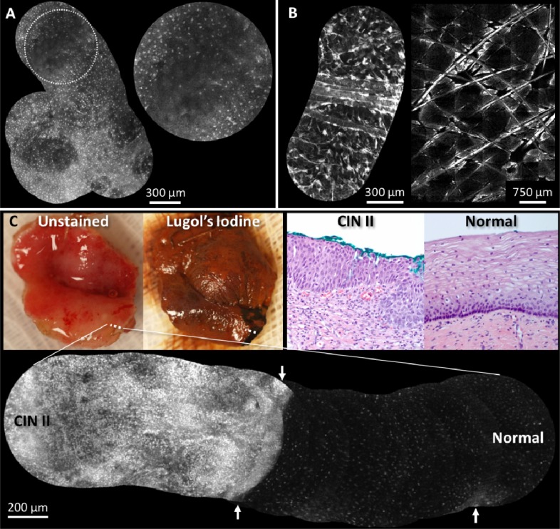
Fig. 4.
Real-time video mosaics (see Media 4 (688.4KB, AVI) ) from (A) normal in vivo oral tissue, (B) normal in vivo epidermis, and (C) pre-cancerous ex vivo cervical tissue. These results show that the algorithm correctly aligns and combines morphological features of several cell types and anatomical locations. In (A) the distribution of normal squamous epithelial cells is seen in the gingiva and in (B, left) features such as hair and keratin sheaths are visible. An image mosaic (B, right) from a benchtop confocal microscope obtained during the same imaging session reveals similar morphologic features as (B, left). The normal/abnormal junction of ex vivo cervical tissue is shown (C), where the white dotted line indicates the path of the mosaic. Lugol’s iodine was used to help visualize the extent of abnormal tissue; dark brown staining indicates normal mucosa. Corresponding histopathology from inside and outside the normal region is shown (images labeled “Normal” and “CIN II,” respectively). The mosaic also shows that it can be useful to display a large mosaic even with misregistered/blurred frames (white arrows).

