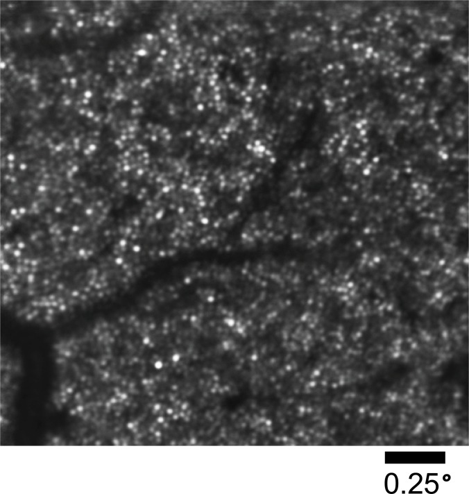
Fig. 10.
Image of the cone photoreceptor mosaic of an emmetropic eye at 4° eccentricity from the fovea. The image is an average of 300 stabilized frames from a single 10-second stabilized video. The bright white circles are individual cone photoreceptors, while the dark shadows extending from the lower left corner into the center of the image are blood vessels.

