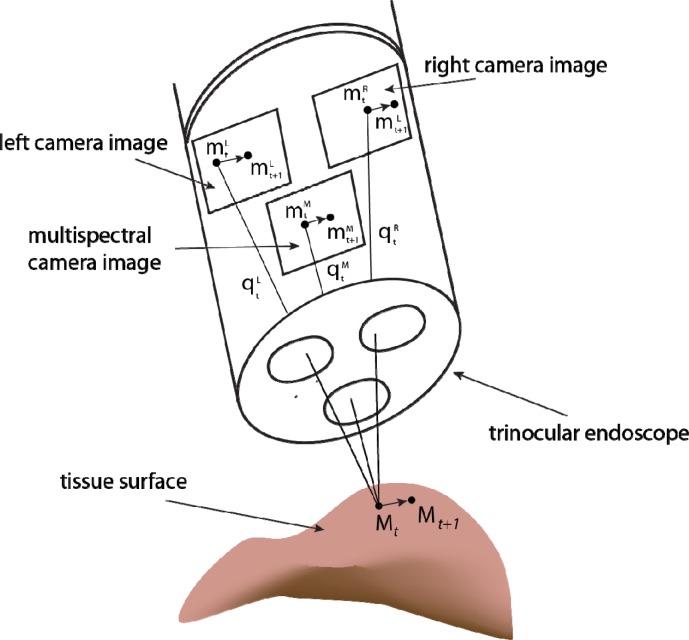
Fig. 2.
Illustration of trinocular endoscope imaging geometry. Geometric calibration of the system means that image points in the left and right white light images can be used to triangulate the 3D position of points on the tissue surface. These can then be reprojected into multispectral image coordinates. We track the motion of points in the white light images as these have consistent light appearance and we use the reprojection capability of the calibrated system to maintain a track of the respective region in the multispectral image.

