Figure 9.
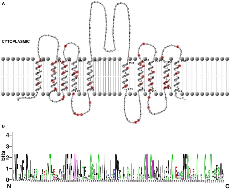
Structure of Arabidopsis thaliana PILS proteins. (A) Predicted topology of A. thaliana PILS5 protein. The prediction was done by HMMTOP 2.0 (Tusnády and Simon, 1998, 2001) and visualized by TMRPres2D (Spyropoulos et al., 2004). Conserved amino acids in all seven PILS proteins are marked in red. (B) Sequence logos generated by WebLogo (Schneider and Stephens, 1990) representing a ClustalW multiple sequence alignment (Larkin et al., 2007) of 109 amino acids from N-terminal region of A. thaliana PILS proteins (exons 2–4). Note the PILS sequence conservation at the highest, single symbol positions.

