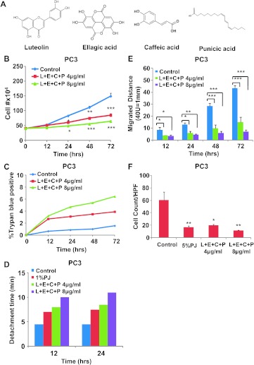
Figure 1.
L + E + C + P inhibits growth, stimulate adhesion, inhibits migration, and inhibits chemotaxis toward SDF1α of hormone-independent prostate cancer cells. (A) The chemical structure of L, E, P, and C. (B) PC3 cells were treated with four PJ components L + E + C + P at 4 and 8 µg/ml and counted for increasing times after initiation of treatment. Controls represent no treatment. The medium containing L + E + C + P was changed daily. (C) PC3 cells were treated with L + E + C + P at 4 and 8 µg/ml, and the percentage of dead cells was determined by Trypan blue staining at the indicated time points. (D) PC3 cells were plated on gelatin-coated dishes, and 24 hours later, the medium was changed and the cells were treated with L + E + C + P at 4 and 8 µg/ml. We tested for adhesion to the substrate at 12 and 24 hours after initiation of treatment by recording the time it took for trypsinization to remove all of the cells from the dish. Control represents no treatment. The reason for not presenting statistical significance is because the loss of adhesion is very similar from culture to culture and it occurs rapidly when the cells begin to detach. Within each experiment, the times of trypsinization were the same within 1 minute for each specific treatment. (E) PC3 cells were treated with L + E + C + P at 4 and 8 µg/ml for 72 hours and the distance migrated by the cells from the wounded edge to the leading edge was measured at the indicated time points. Controls represent no treatment. The medium containing the components was changed daily. (F) PC3 cells were allowed to attach to the top of the filter of the chemotaxis assay chambers for 4 hours and then treated with L + E + C + P at 4 and 8 µg/ml for 12 hours. At this time, 100 ng/ml SDF1α was introduced into the lower chamber and the cells found on the bottom of the filter were counted 3.5 hours later. Control had no treatment. The number of cells found on the underside of the filter was counted 3.5 hours later. Bars represent SEM. ***P < .001; **P < .01; *P < .05.

