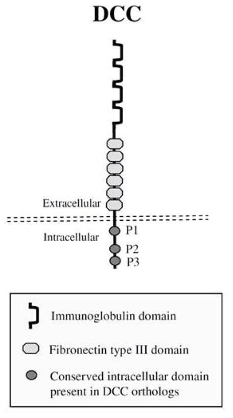
Fig. 1.

DCC protein structure. A schematic representation of DCC with the location of the immunoglobulin, fibronectin type III, transmembrane, as well as the conserved intracellular P1, P2, and P3 domains noted. The structure depicted is adapted from reference [7].
