
Fig. 3.
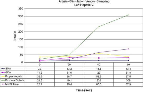
Left hepatic vein insulin concentrations after intra-arterial calcium injection. Injections of the superior mesenteric artery (SMA), gastroduodenal artery (GDA), and proper hepatic artery do not show any suspicious areas. However, the increase in insulin concentration after injection into the proximal and mid splenic arteries help localize this lesion to the tail of the pancreas. (Courtesy of National Institutes of Health, Bethesda, MD.)