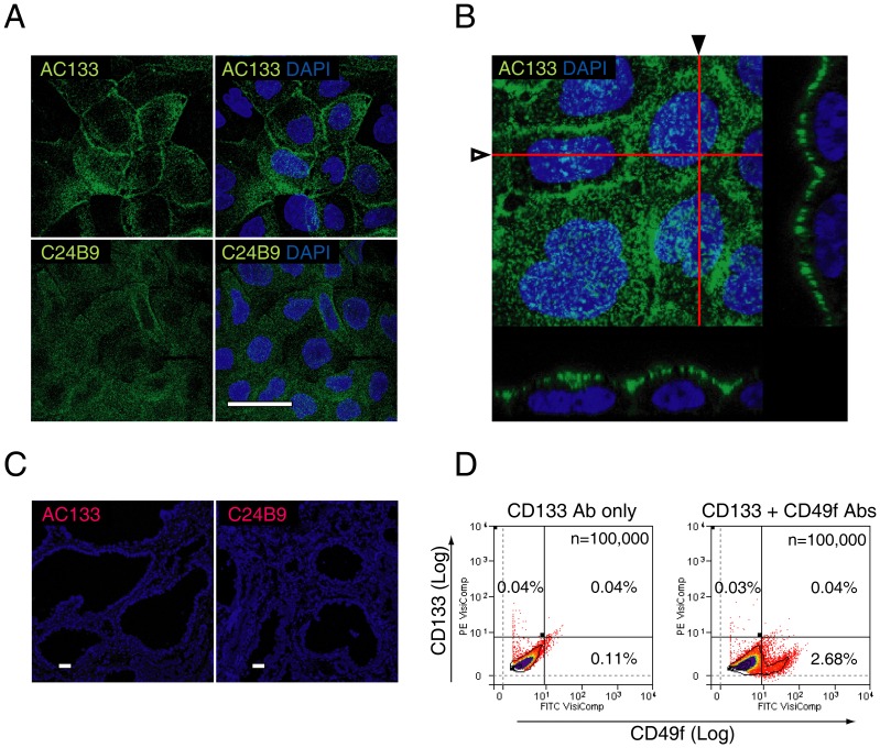
Figure 4. CD133 expression in frozen prostate tissue sections.
Validation of CD133 (clones AC133 and C24B9) antibody specificity and expression in the human prostate. [A] Punctate expression was shown on the cell surface of Caco-2 cells for clones AC133 and C24B9, as described previously [21]. [B] Orthogonal sectioning following three-dimensional reconstruction of 150 slices (red lines marked by hollow and solid arrowheads indicate the x-z planes shown below, or to the right of the confocal image, respectively) indicate CD133 expression only along the apical border of the plasma cell membrane [21]. [C] Immunohistochemical expression of AC133 and C24B9 in prostate tissue. Each frozen tissue section measured 10×10 mm in cross-sectional area. Following examination of 20 slides each from 5 patients, we found no cell with definitive membrane expression. [D] Flow cytometric co-expression analysis of CD133 and CD49f. A representative flow cytometric analysis of 3 patients shows that CD133+ cells and CD49f+ cells are mutually exclusive populations, with no significant increase in the percentage of cells within the CD49f+/CD133+ cell gate compared to control. Scale bar = 20 µm.

