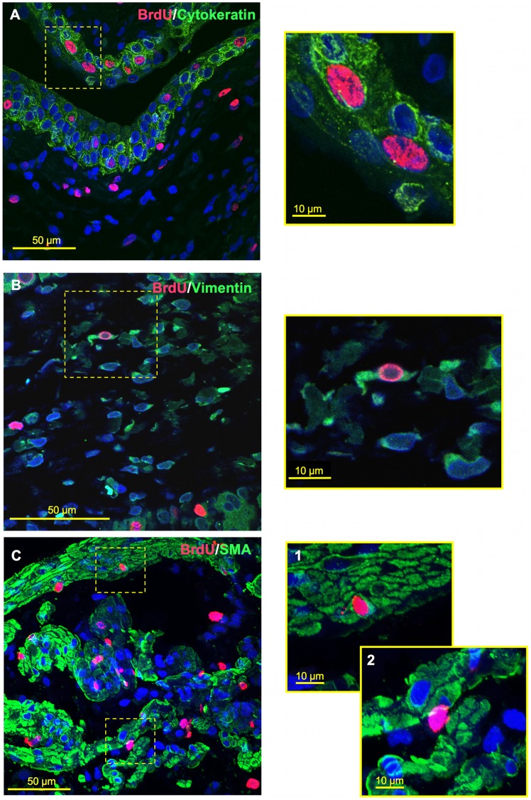
Figure 6. Co-labeling of BrdU expressing cells with cell-specific markers for the urothelium, MP and LP.
Confocal z-stack reconstruction imaging was performed at 600x magnification, where offset pictures are digitally zoomed. (A) BrdU-cytokeratin co-labeling within urothelium at 3-days post-STC. (B) BrdU-vimentin co-labeling with LP at 3-days post-STC. (C) 7-days post-STC BrdU-SMA co-labeling was also observed within the MP (C-1), but was relatively rare. BrdU-labeled cells within the MP were more commonly observed between smooth muscle cells as well as smooth muscle bundles (C-2).

