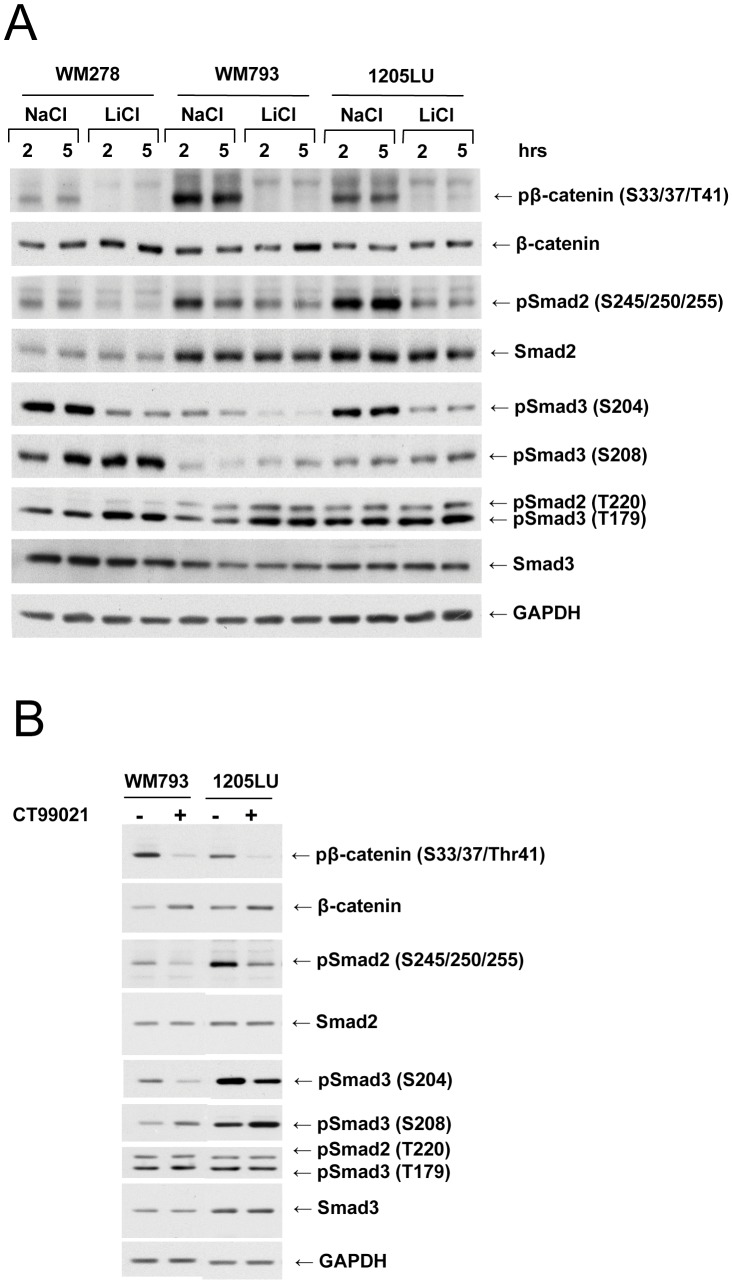
Figure 1. Smad linker phosphorylation involves GSK3 in human melanoma lines. A.
Inhibition of Smad2 and Smad3 linker phosphorylation in the presence of LiCl. 24 hours post seeding, WM278, WM793 and 1205LU human melanoma cell lines were serum-starved for about 16 hours, before the addition of 50 mM LiCl for 2 and 5 hours. Cells left with 50 mM NaCl for 2 or 5 hours were used as controls. Whole cell extracts were then prepared. Immunoblots were performed with antibodies against: Phosphorylated β-catenin (pβ-catenin); total β-catenin; Both phosphoSmad3 (Thr179) and phosphoSmad2 (Thr220); phosphoSmad2 (Ser245/250/255); phosphoSmad3 (Ser204); phosphoSmad3 (Ser208); Smad2 and Smad3; GAPDH. B. Inhibition of Smad2 and Smad3 linker phosphorylation in the presence of the specific GSK3 specific inhibitor, CT99021. 24 hours post seeding, WM793 and 1205LU cells were serum-starved for about 16 hours, and incubated in the absence (−) or presence (+) of 2 µM of CT99021 for two hours. Immunoblots were performed as in A. p: Phosphorylated. S: Serine; T: Threonine.

