
Figure 3.
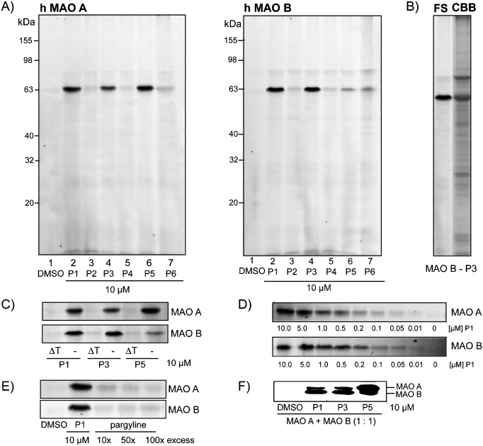
Fluorescent SDS-PAGE analysis of the labeling of monoamine oxidases A and B in vitro. A) Screening of alkyne probes P1–P6 with MAO A and MAO B. B) Comparison of fluorescence scanning (FS) and Coomassie Brilliant Blue (CBB) staining of the labeling of MAO B by the probe P3. C) Labeling of MAO A and MAO B after heat denaturation (6 min at 96 °C) (right) and without heat denaturation (left) of the enzyme. D) Concentration-dependent labeling of MAO A and MAO B by probe P1. E) Competitive labeling of MAO A and MAO B with pargyline and probe P1. F) Labeling of mixture of both MAO isoforms with probes P1, P3, and P5.