Figure 2.
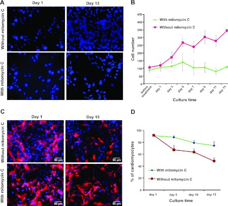
Treatment with mitomycin C inhibits cardiac fibroblast proliferation (A, B) and increases the percentage of cardiomyocytes in rat cardiomyocyte culture (C, D). A) Fluorescent images of a rat cardiomyocyte culture stained with DAPI on d 1 and 13 after seeding. B) Growth rate of cells was measured by counting DAPI-positive cells in cardiomyocyte culture at different time points. C) Immunofluorescent staining analysis of troponin I expression in rat cardiomyocyte culture on d 1 and 15 after treatment with or without mitomycin C. Blue staining indicates nuclear signals; red staining indicates troponin I signals. Scale bars = 50 μm. D) Percentage of troponin I-positive cardiomyocytes in rat cardiomyocyte culture over time after treatment with or without mitomycin C. Data are expressed as means ± sd (n=3).

