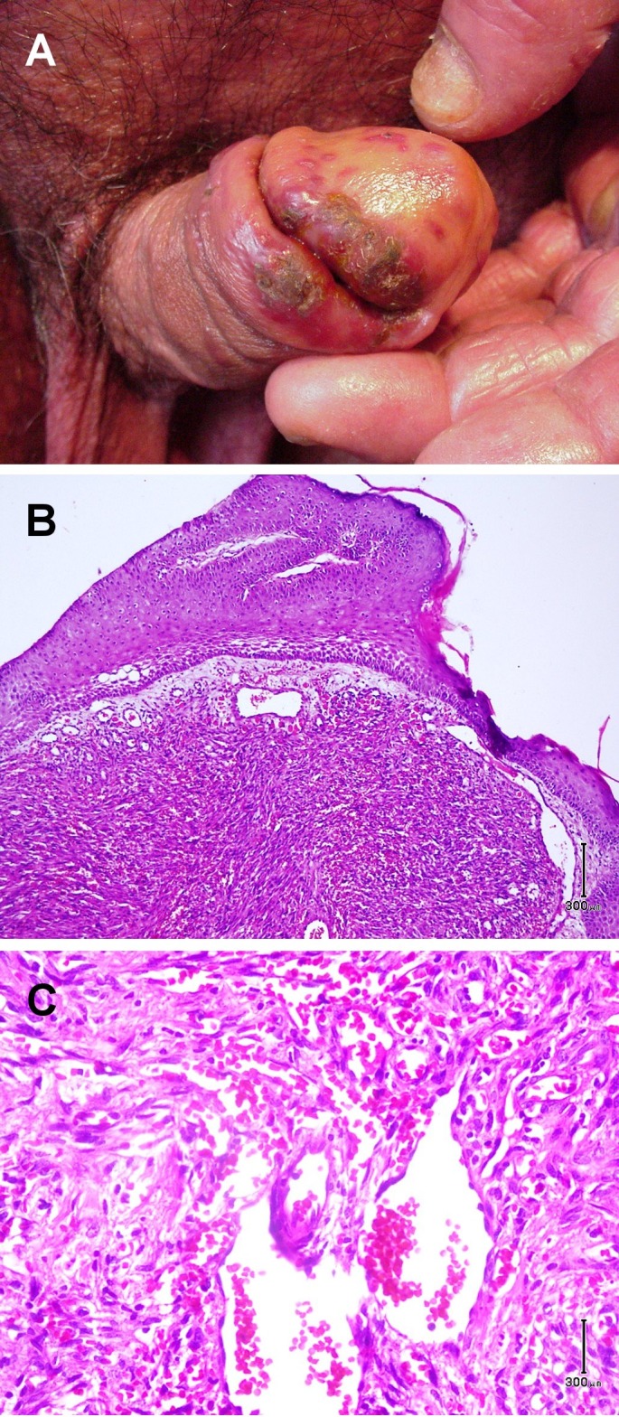
Figure 2.

(A) Multiple red, violaceous papules coalescing to an impetigenized plaque with yellow-brown scale crust on the glans of penis and sulcus corona. (B-C) Histological view of nodular vascular proliferation with spindle slit-like lumina, dilated blood vessels and hemorrhage. (H&E 40 and 100).
