
Fig. 1.
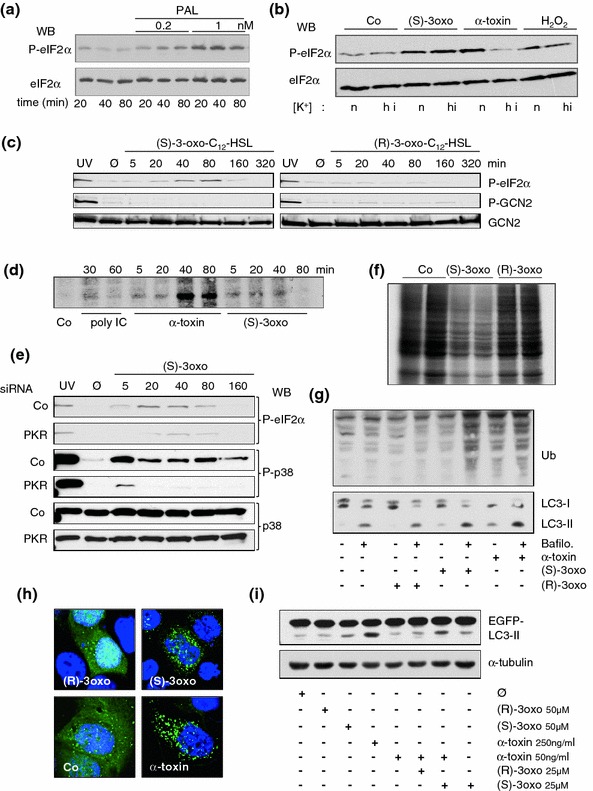
A quorum-sensing hormone of P. aeruginosa impacts translation and autophagy. Materials and methods employed here have been published previously [59, 88]. Metabolic labeling was performed as described by [89]. In the figure, (S)-3oxo and (R)-3oxo denote (S)-3-oxo-C12-HSL and (R)-3oxo-C12-HSL, respectively. a Western blot for (p)-eIF2α with whole cell lysates (HaCaT) treated with palytoxin (PAL) as indicated in the figure. b Western blot for (p)-eIF2α with whole cell lysates (HaCaT) treated with the indicated compounds in the presence of normal concentrations of potassium (n), or in media with high concentration of potassium (hi) [59]. c Western blot for p-eIF2α, p-GCN2, and GCN2 after treatment of HaCaT cells with the P. aeruginosa quorum-sensing hormone (S)-3-oxo-C12-HSL or the control compound (R)-3-oxo-C12-HSL for the indicated times. As expected, (R)-3-oxo-C12-HSL fails to cause phosphorylation of eIF2α. Note that GCN2 is not phosphorylated in response to either lactone; UV served as a positive control. Untreated cell samples (media alone) are denoted Ø. d Autoradiographic detection of P32-PKR in samples of HaCat cells treated for the indicated times with the compounds denoted underneath the panel. Co: medium alone. e Western blots for p-eIF2α, p-p38, or p38 with straight Cos7-cell lysates obtained 48 h following transfection with siRNAs and subsequent treatment with S-3-oxo-C12-HSL. f Autoradiography of an SDS-Gel visualizing incorporation of S35-Methionine into newly synthesized proteins. Note marked inhibition of protein synthesis in cells treated with S-3-oxo-C12-HSL. g Western blots for ubiquitination and LC3I/II with lysates of HaCat cells treated as indicated in the figure for 3 h. h Fluorescence microscopy images of HaCat cells transfected with EGFP-LC3 and treated for 3 h with compounds indicated in the figure. Note redistribution of diffuse green fluorescence signal into dots in cells treated with α-toxin, or S-3-oxo-C12-HSL. i Western blot for GFP with lysates of HaCat cells transfected with EGFP-LC3 treated as indicated in the figure. The untreated cell sample (media alone) is denoted Ø. Loading control with α-tubulin. Combination of (S)-3-oxo-C12-HSL and α-toxin led to a significant accumulation of LC3II