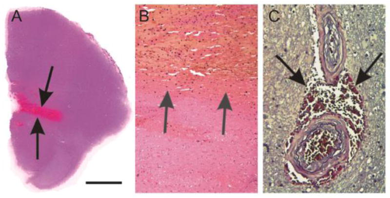
Fig. 2.

Hemorrhage and microbleeds. A, B: Cerebral hemorrhage in the pons (arrows in A). At higher magnification level (B) it becomes evident that blood clots (arrows) displace the vital brain parenchyma. C Microbleed in the basal ganglia. The microbleed, thereby, represents a hemorrhage restricted to blood extravasation into the perivascular space (arrows) without tissue damage and displacement. Note the two arteries associated with the microbleed exhibit pattern of SVD with a concentric intima proliferation and partial fibrosis of the vessel wall.
Calibration bar in A corresponds to A = 4500μm, B = 226μm, C = 85 μm. A–B: Hematoxylin & Eosin staining; C: Elastica van Gieson staining.
