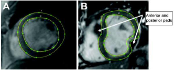
Figure 2.

Contoured short-axis cardiac MR images before (A) and after (B) surgical implantation of the Coapsys device. The contours from multiple short-axis images covering the LV from apex to base were used to create patient-specific 3D finite element models.
