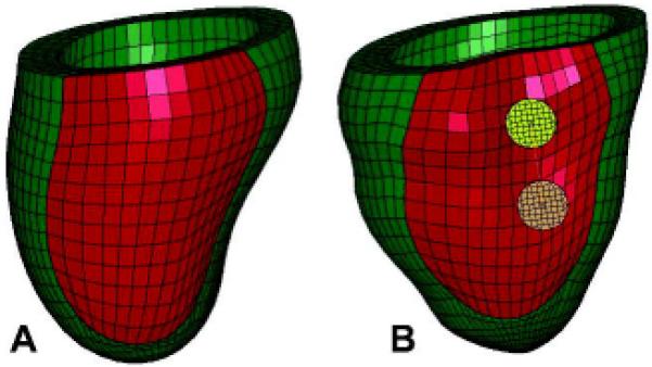
Figure 3.

Two views of the unloaded post-Coapsys finite element model showing (A) the PRE-OP model and (B) the COAPSY SUBACUTE model showing the double pad side of the device. Infarct regions are colored with red while the remote regions are colored with green.
